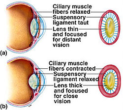
Ciliary Body
|
A circular band of tissues at the front of the Choroid that supports the Lens. It contains Ciliary Muscles that adjusts the curative of the Lens.Ciliary Muscles determines the shape of the Lens. When the muscle contracts, the lens is rounder, thicker, the focal length decreases and more convex and light rays from a near object are sharply focused on the Retina. When the muscle relaxes, the Lens is flatter, the focal length increases and less convex and the light rays from a distant object are sharply focused onto the Retina. This can be described as Antagonistic (when one sewt contracts, the other relaxes)
the information on this section was obtained from: thevisibleworld |
|
Photoreceptors

|
-
Rods are responsible to detect light in dim light (black and white) enabling night vision. They contain a pigment in rods called Rhodospin which is light sensitive. If exposed to bright light then it will be bleached. Rhodospin enables a person to see in the dark.
-
Cones are responsible to detect coloured light. There are green, blue and red cones. Red is used to respond to long wavelengths. Blue is used to respond to short wavelengths and Green is used to respond to medium lengths. Cones are closely paccked to allow the perception of details but cannot work well in dim light.
the information on this section was obtained from: thevisibleworld |
|
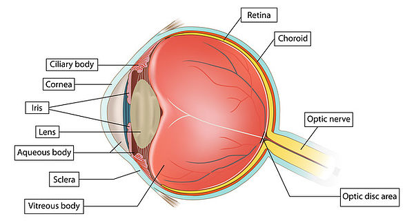
Iris
Is the coloured part of the eye; eg. Asians and the coloured race generally have a brown iris and the Caucasions/Whites generally have blue or green iris. The central opening is the Pupil of the eye.
Pupil
Is the round opening at the centre of Iris, through which light passes into the eye. The pupil
acts like a camera’s aperture; it increases or reduces in size to adjust for the optimum amount
of light needed for clear vision.
The pupil increases in size in the dark and reduces in size in the light.
Cornea
Is the round transparent layer at the front of the eye; through which we can see the iris. The cornea helps to refract and to focus light onto the retina.
This is the part that we can transplant from a donor eye to a recipient eye. Transplantation of a whole eye-ball is not possible at present.
Lens
Is a transparent round structure placed just behind the iris. It helps to refract and to focus light onto the retina.
Once the lens become cloudy with aging or due to secondary cause, the amount of light entering the eye will be reduced and vision will be blurred.
Retina
Is a light-sensitive layer of tissue lining the inner wall of the eye ball. It contains millions of photoreceptors – ‘rods and cones’ that are essential for perception of sharp images.
Macula & Fovea
Macula is the centre part of the retina; which contributes to the sharp image on direct gaze/central vision. The centre of it is called Fovea.
Damages on the macula will affect our central vision. Central vision is important for us to be able to perform fine task, to recognize faces and to read.
Optic Disc and Optic Nerve
Optic disc is the centre point where all nerve fibres carrying messages from the photoreceptors in the retina converge to form the Optic Nerve. We have approcimately 1.2 millions of nerve fibres in one eye.
Eye diseases or trauma that affect the Optic Disc and Optive nerve can cause blindness.
Sclera
Is the white outer protective coat of the eye. It is covered on its front surface by a thin layer of tissue called conjunctiva.
Conjunctivitis is a common eye surface disease causing red eyes.
Why your eyes are so important?
- Eyes provide the most important sense of 5 senses that human beings possess – that is your vision. Your vision contributes about 75% of all your senses.
- A person who has lost one eye lost 25% of his functional capacity; A person who has lost both eyes would loss 75% of his functional capacity.
- A blind person has significant impairment of quality of life. He lives a life with no colours; everything in life would appear dull.
- A blind person is more prone to depression compares to a man with normal sight.
- A blind person is 3-10 times more prone to injury/accidents.
- A blind person is a burden to his family and society; in terms of social, psychology and economy.
|